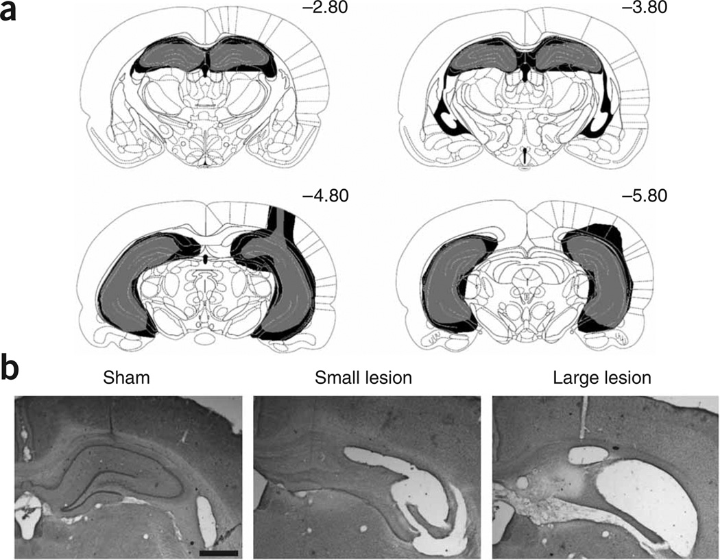
Figure 1.
Rats in the experimental group were given excitotoxic lesions of the complete hippocampus. (a) The largest lesion (in black) and smallest lesion (in gray) were selected from all animals included in the study, and are shown here according to a rat brain atlas15 (Supplementary Results online). (b) Representative examples of the smallest and largest lesion are shown. Scale bar, 1 mm. These experiments were conducted in accordance with procedures outlined by the Animal Care and Facilities Committee at Rutgers University.

