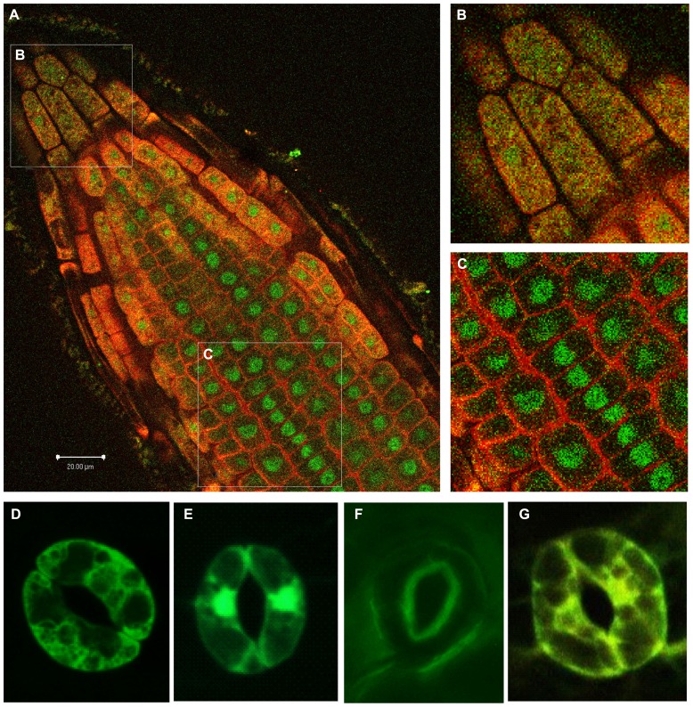
FIGURE 6.
Distinctly different 14-3-3 localizations determined by confocal microscopy of Arabidopsis. Isoform-specific antibodies that have been directly labeled with either Alexa-488 or Alexa-568 and used for in situ labeling. 14-3-3 λ (green) predominates in the nucleus while 14-3-3 ε (red) predominates in the cytoplasm and plasma membrane. A single longitudinal optical slice of a root tip is shown in (A). (B,C) Provide closer views of the boxed sections of the root in (A). The root tip (B) exhibits an even distribution of 14-3-3-λ and 14-3-3-ε throughout the cells, and a section father back in the root (C) exhibits a clear partitioning of 14-3-3-λ into the nucleus and 14-3-3-ε to the plasma membrane/cell wall. Guard cell subcellular localization of four GFP-tagged 14-3-3 isomers is shown in the bottom row: ε (D), κ (E), λ (F), and φ (G).

