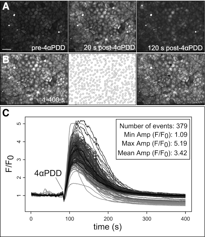
Fig. 4.
Automated analysis of 4αPDD-evoked Ca2+ events in rat pulmonary microvascular endothelial cell (RPMVEC) monolayers. Automated ROI analysis was applied to confocal image sequences of cultured RPMVEC monolayers loaded with Fluo-4 Ca2+ indicator dye and stimulated with the phorbol ester 4αPDD. A: the addition of 4αPDD stimulated a widespread rise of intracellular Ca2+ levels at multiple cellular sites, shown at selected prestimulated, maximally responding (20 s), and poststimulated (120 s) time points. Scale bar represents 10 μm. A time lapse accumulate image (B) shows cell sites that responded to treatment within the time course (left), corresponding to circular ROIs that were automatically assigned to active sites by our algorithm (middle), and an overlay of the accumulate image and detected ROIs (right). Arrows in the overlay (right) indicate cells with single (bottom) and multiple ROIs (top). Measurements of average intensity within each ROI are shown for the time course of the image sequence (C) with the event number and mean amplitude value and range summarized in the inset.

