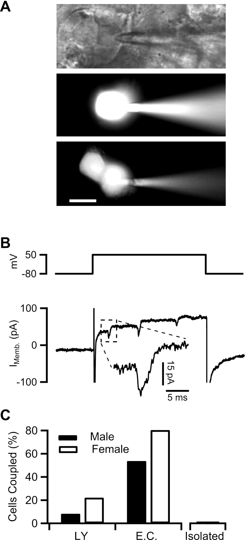
Fig. 1.
Gap junction coupling in the adrenal medulla. A, top; a bright-field image of a chromaffin cell in situ patched in whole cell configuration. Pipette solution contained 1 mM LY dye. Middle and bottom: the cells were held at −80 mV. Cells are shown 20 min after break-in to allow Lucifer yellow (LY) diffusion into the cell (scale bar = 10 μm). B, top: cell from A, middle, was held at −80 mV and delivered a 150-ms duration depolarizing step to +50 mV. Bottom: evoked membrane current is plotted. B, inset: echoing action potential (Imemb) from a neighboring unpatched cell. C: data were collected from LY dye diffusion and electrical stimulation experiments as represented above and are plotted as percentage of cells that are coupled. Sample sizes were as follows: for LY diffusion, n = 30 cells (12 male and 18 female) and for electrical coupling (EC), n = 63 cells (29 male and 34 female). Data from isolated cells are provided as a negative control (n = 10 cells).

