Fig. 5.
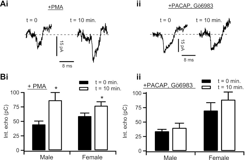
PACAP enhances electrical coupling through a PKC-dependent mechanism. Ai: electrical echoes were measured in chromaffin cells, as in Figs. 1–4, before (t = 0 min) and after (t = 10 min) focal and bulk perfusion of 100 nm PMA. Representative electrical echoes from each time point are shown. Aii: protocol was repeated in male mouse chromaffin cells (n = 10 echoes) and female mouse chromaffin cells (n = 22 echoes). Data from each PMA-stimulation time point were separated by gender and plotted as means ± SE. Male: *statistical significance at P = 0.025; female: *statistical significance at P = 0.048. Bi: an adrenal tissue slice was pretreated for 5 min with focal and bulk-perfused Gö6983 (100 nM) and then stimulated with focally perfused 1 μM PACAP for 10 min. Representative electrical echoes from the 0- and 10-min time point of PACAP stimulation are shown. Bii: protocol was repeated in male mouse chromaffin cells (n = 10 echoes) and female mouse chromaffin cells (n = 7 echoes). Data from each PACAP + Gö6983 stimulation time point were separated by gender and plotted as means ± SE. Male: P = 0.344; female: P = 0.306.

