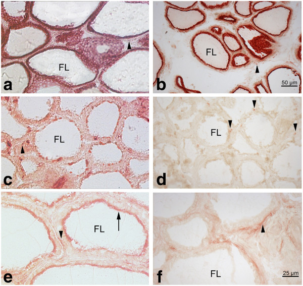
Figure 1 .
Detection of protease activity with synthetic substrates by histochemistry (red) in porcine (a, c) and bovine (b, d) thyroid tissue. Activities of perifollicular cells (endothelial cells, fibroblasts and C-cells) for the respective proteases are indicated by arrowheads. a, b: Activity of dipeptidyl peptidase II is seen intracellularly in thyrocytes of both species. c: In porcine thyroids activity of dipeptidyl peptidase IV is seen in some follicle cells. d: In bovine thyroids, follicle cells show no activity for dipeptidyl peptidase IV substrate. e: Activity of aminopeptidase N is seen at the apical part of the cells in porcine follicular thyrocytes (arrow), whereas in bovine thyroids (f) activity of aminopeptidase N is restricted to endothelial cells the perifollicular stroma below the follicular epithelium (arrowhead). FL: follicle lumen.

