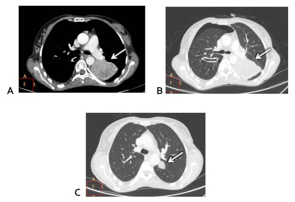
Figure 2.
Computed tomography scan evaluation. (A) Baseline: great metastatic nodular lesion on the left lung (white arrow). (B) Baseline: lung parenchymal windows showing the great lesion on the left and a drainage tube for thoracentesis with residual pneumothorax (white arrow). (C) Week 12 under treatment: reduction of the great lung lesion with small residual nodule (white arrow).

