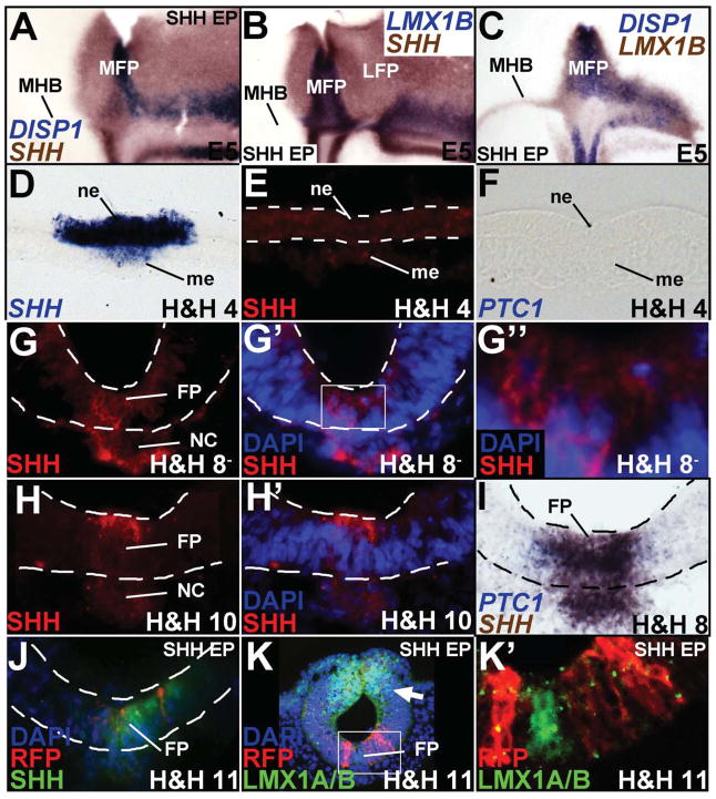
Figure 2. SHH misexpression is sufficient for LFP, but not MFP induction.
(A–C, flattened wholemounts, oriented as in Fig. 1C; D–K′, cross-sections). (A–C) Unilateral SHH misexpression is sufficient to convert the entire right half of the midbrain into SHH+ LFP (A, B), but can only specify an LMX1B+/DISP1+/SHH+ MFP along the MHB (A–C). (D–F) SHH mRNA (D) but no SHH protein (E) or PTC1 transcripts (F) at H&H 4. Sections in D–F are drawn from presumptive neural plate anterior to Hensen’s node. (G–H′) SHH protein (red) at H&H 8− (G–G″) and H&H 10 (H–H′). G′ and G″ show the micrograph in G with DAPI staining. The boxed area in G′ is magnified in G″ to show a SHH+ cell (arrowhead) and a SHH-negative cell (arrow). H′ shows the image in H with DAPI staining. (I) SHH and PTC1 mRNA at H&H 8. (J, K′) H&H 4–6 SHH-ires-RFP electroporation (red) is sufficient for ectopic, non-autonomous SHH (J, green) induction and dorsal LMX1A/B suppression (arrow, K), but it is not sufficient for LMX1A/B (green) induction ventrally (K, K′). Boxed area in K is magnified in K′ to show the absence of LMX1A/B induction in or around SHH -ires-RFP+ cells. Please note that the sections shown in G–K′ are drawn from the rostrocaudal midpoint of the midbrain.

