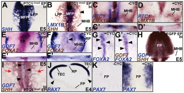
Figure 3. SHH is necessary for MFP induction.
(A–B) Broad, bilateral misexpression of PTC1?Δloop2 disrupts the SHH+/LMX1B-negative LFP (B, arrowhead), but does not affect the SHH+/LMX1B+ MFP. Control for 3B can be seen in Figure 4E. (C, D) H&H 3 explants treated with either vehicle (−CYC) or cyclopamine (+CYC) for 24 hours demonstrate that HH blockade suppresses PTC1 expression in LFP, but does not affect LMX1B expression in the MFP. (C′, D′) Cross-sections of the wholemounts shown in C and D. (E, E′) Control explants in flattened wholemount (E) and cross-sectional (E′) view displaying GDF7 (RP) and FOXA2 (FP) expression. (F, F′) Cyclopamine treated explants displaying the loss of FOXA2 and the ectopic induction of GDF7 at the ventral midline (arrowhead). Note that compared to E, E′, the roof plate (RP) expression of GDF7 is expanded in F, F′. (G, G′) Cyclopamine treated explants with mild HH blockade showing ectopic expression of GDF7 (arrowheads) in the ventral midline whereever FOXA2 expression is abolished. Note that G and G′ show the same explant before and after GDF7 staining. Control for G, G′ is shown in Fig. E. (H) Control brain demonstrating that GDF7 is not expressed in the SHH+ FP or MHB. See also, 1N; 7G, H for controls. (I) Bilateral HH blockade results in a disruption of the LFP (red arrows) and a conversion of the MFP into GDF7+ RP (arrowheads). (J) Midbrain cross-section demonstrating that PAX7 expression is absent from the RP and ventral midbrain, but present throughout dorsal midbrain (TEC). (K, L) Control (K) and cyclopamine treated explants (L) shown in wholemount view demonstrating that the ventral expansion of PAX7 into ventral midbrain (K) excludes the ventral midline. The PAX7-negative region between the arrowheads in J was flattened in K, L.

