Figure 7.
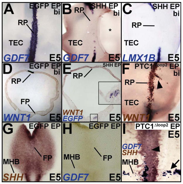
(A–E) Top down (A–C) and cross-sectional views (D, E) demonstrating that compared to EGFP-electroporated controls (A, D), RP induction is blocked by ventral electroporations of SHH. Inset in E shows magnified view of the boxed region and demonstrates the location of SHH misexpression by EGFP transgene expression. * in B marks a hole cut in dorsal midbrain for irrigation. (F) Ventral PTC1Δloop2 misexpression results in ectopic RP induction (arrowhead). (G–I) Midbrain flatmounts showing that unlike controls (G, H), PTC1Δloop2 misexpression results in reduced SHH expression accompanied by ectopic GDF7 expression in both the ventral midline (arrowhead) and the MHB (arrow).

