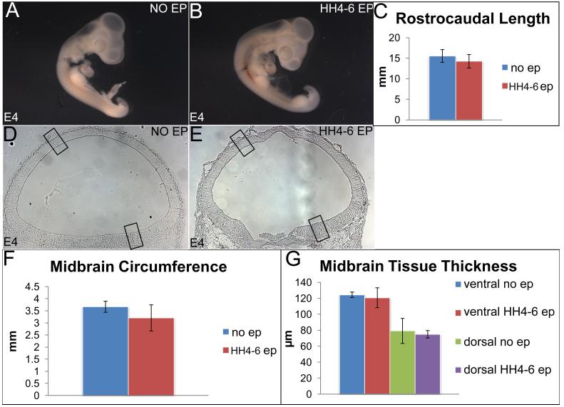
Figure 3. Early electroporations do not affect the size of the embryo, midbrain size and morphology.
(A-C) Sagittal view of un-electroporated (A) and early-electroporated (B) embryos showing no significant differences in their rostrocaudal length. (C) Quantitation of A, B (rostrocaudal length, un-electroporated: 15.55 +/− 0.525mm; early-electroporated 14.27 +/− 0.945 mm; p=0.525. (D, E) Un-electroporated (D) and early-electroporated midbrains (E) showing similar sizes and tissue morphology. (F, G) Quantitative data demonstrating that midbrain circumference (F) and ventricular-pial thickness (G) do not differ in un-electroporated and electroporated embryos. (Circumference: un-electroporated: 3.67 +/− 0.229 mm; early electroporation: 3.20 +/− 0.540 mm; p= 0.495; ventral thickness: controls 124.38 +/− 3.35 μm; electroporated: 120.68 +/− 12.44 μm; p=0.798; dorsal thickness: controls: 79.11 +/− 15.66 μm; electroporated 79.94 +/− 4.60 μm; p=0.819).

