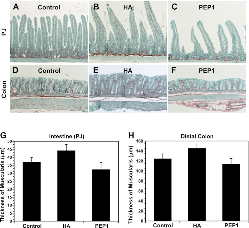
Fig. 9.
Sirius red staining for total collagen (red) and fast-green staining for noncollagenous protein (green) in small intestine (PJ, A–C) and DC (D–F). Collagen staining in the lamina propria shows changes in intestinal and colonic morphology due to HA or PEP-1 treatment confined to the mucosa. Average villus height (intestine) and crypt depth (intestine and colon) were greater in HA-treated mice than controls, while average villus height and crypt depth were smaller in PEP-1-treated mice than controls. Original magnification ×100. G and H: there was no significant effect on thickness of the muscularis with HA or PEP-1 treatment in intestine and colon. Values are means ± SE for 4 mice per group.

