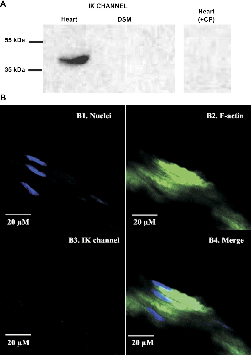
Fig. 4.
Western blot and immunohistochemical detection of IK channel showing the lack of IK channel protein expression in native human whole DSM tissues. A: protein expression for IK channels was detected by Western blot in human heart protein medley (positive control) but not in native human whole DSM tissue. The immunoreactive band in human heart protein medley was eliminated by +CP. Experiments were conducted in 3 separate Western blot reactions using protein isolated from 4 patients. B: IK channel protein was not detected in mucosa-free human whole DSM tissue following immunohistochemical reaction using IK channel-specific antibody. Cells' nuclei are shown in blue (B1); F-actin is shown in green (B2). Lack of red staining in B3 indicates no expression of IK channel. The merged images of the nuclei and F-actin are illustrated in B4. Images were captured with a Carl Zeiss LSM 510 META confocal microscope. Experiments were conducted on tissue samples isolated from 4 different patients.

