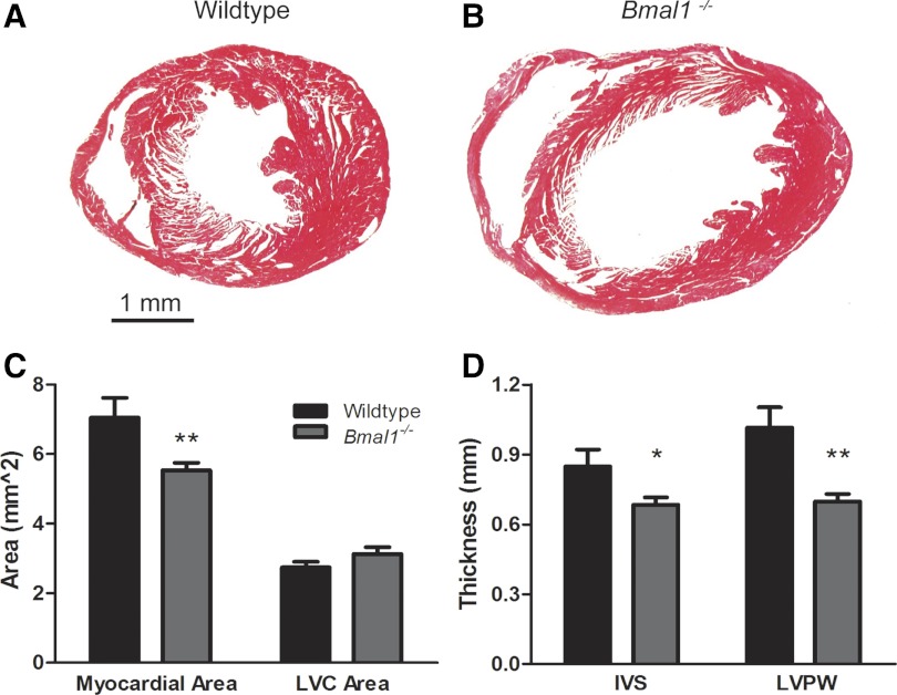
Fig. 3.
Thinning of the myocardial walls and dilation of the ventricular cavity in 36-wk-old Bmal1−/− mice. Representative Masson's trichrome-stained myocardial sections at the level of the papillary muscles, show enlargement of the ventricular cavity and wall thinning in the 36-wk-old Bmal1−/− myocardium (B) compared with an age- and sex-matched WT control myocardium (A). Graphs of combined data show a decrease in the area of myocardium (C) and the thickness of both the IVS and LVPW (D). LVC, LV cavity. n = 5 WT and 10 Bmal1−/− mice. *P < 0.05; **P < 0.01.

