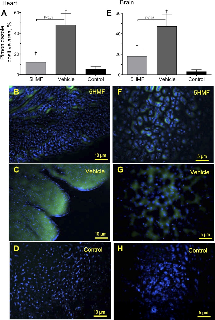
Fig. 7.
Brain and heart pimonidazole binding to hypoxic areas in mice. C57BL/6 mice treated with 5HMF or vehicle were exposed to an identical hypoxic protocol as hamster. Pimonidazole preferentially binds to hypoxic cells, so detection of pimonidazole adducts using monoclonal antibodies can serve as a method for measuring tissue hypoxia. Pimonidazole stained areas in the heart (A–D) and brain (E–H) sections after hypoxia. Superimposed images [hearts (B–D) and brains (F–H)] indicate clear colocalization of pimonidazole (green) and Hoechst (blue). Control animals received pimonidazole but were not exposed to hypoxia. †P < 0.05 compared with control.

