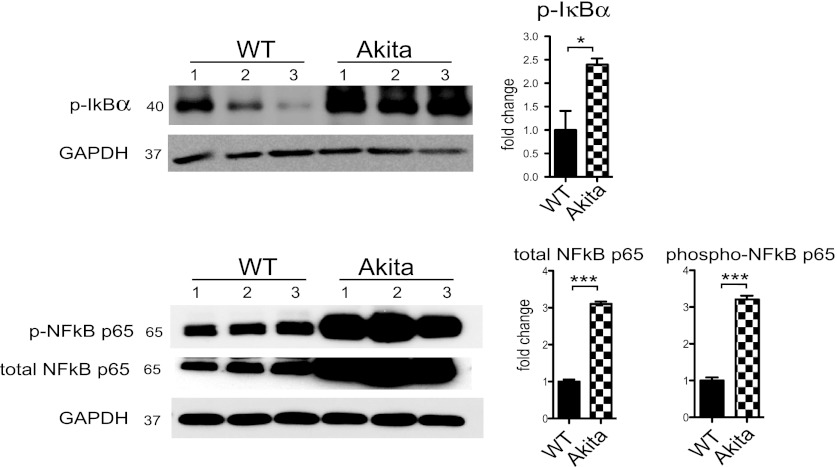
Fig. 7.
Western blot analysis of IκB-α and NF-κB p65 in the Akitains2 whole ventricular tissue. Akitains2 heart showed ∼3-fold elevation of phospho-IκB-α and NF-κB p65 as well as total NF-κB p65. GAPDH was used as loading control. Data shown are means ± SD from at 3 independent experiments; *P < 0.05; ***P < 0.001.

