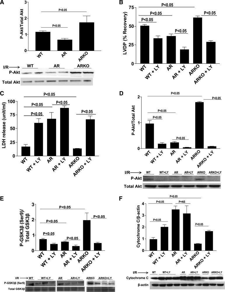
Fig. 4.
p-Protein kinase B (Akt). Western blot analysis for p-Akt in LAD ligation of WT, ARTg, and ARKO hearts (A), determination of myocardial ischemic injury and function, as shown by LVDP (B) and LDH release (C) in WT, ARTg, and ARKO perfused with and without phosphatidylinositol 3-kinase (PI3K)/AKT inhibitor LY-294002. Western blot analysis for p-Akt (D), p-GSK3β-Ser9 (E), and cytochrome c (F) in WT, ARTg, and ARKO hearts perfused with and without PI3K/Akt inhibitor LY-294002. Treatment with LY-294002 decreased p-Akt and p-GSK3β levels in all 3 groups (P < 0.05). Cytochrome c levels were increased in WT and ARKO hearts (P < 0.05), but not ARTg upon perfusion with LY-294002 (n = 4–16 mice/group).

