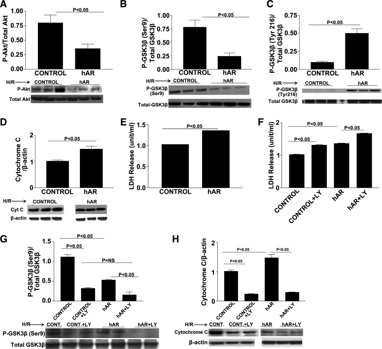
Fig. 6.
HL-1 cardiomyocyte studies. Western blot analysis for p-Akt (A), p-GSK3β-Ser9 (B), p-GSK3β-Tyr216 (C), and cytochrome c (D) in control and hAR-overexpressing HL-1 cells subjected to H/R alone. E: determination of cardiomyocyte H/R injury as shown by LDH release in control and hAR-overexpressing HL-1 cell supernatants. F: LDH release in control and hAR-overexpressing HL-1 cells treated with and without PI3K/Akt inhibitor LY-294002. Western blot analysis for p-GSK3β-Ser9 (G) and cytochrome c (H) in control and hAR-overexpressing HL-1 cells treated with and without PI3K/Akt inhibitor LY-294002. Cells were incubated with LY-294002 (10 μM) or its vehicle control dimethyl sulfoxide (DMSO) for 1 h, followed by 30 min of hypoxia and 1 h reoxygenation (H/R). Treatment with LY-294002 decreased p-Akt levels, decreased inhibition of GSK3β, and increased apoptosis in both control and hAR-overexpressing cells. LDH release in supernatants in both control and hAR-overexpressing cells was increased with PI3K/Akt inhibitor LY-294002 treatment. Error bars not visible. Control: SE ± 0.006; and hAR: SE ± 0.011.

