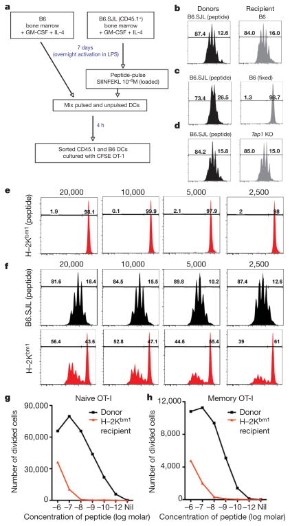
Figure 1. Transfer of peptide-loaded class I molecules between dendritic cells in vitro.
a, Scheme of the experiment. DC, dendritic cell; GM-CSF, granulocyte macrophage colony-stimulating factor. b–d, In-vitro-generated bone-marrow-derived B6.SJL dendritic cells were peptide pulsed and cultured with unloaded bone-marrow-derived B6.GFP dendritic cells (b), fixed B6.GFP dendritic cells (c) or Tap1−/− dendritic cells (d). Dendritic cells were separated by cell sorting and 2 ×10⁴ cells were cultured with CFSE-labelled OT-I cells for 60 h. Representative flow cytometry profiles are depicted. e, H–2Kbm1 dendritic cells were pulsed with peptide before culture with CFSE-labelled OT-I cells. f, Peptide-pulsed B6.SJL dendritic cells were cultured with unloaded H–2Kbm1 dendritic cells. Dendritic cells were then separated and cultured with CFSE-labelled OT-I cells. Representative flow cytometry profiles are shown. The numbers above the plots indicate the number of dendritic cells cultured per well. Bone-marrow-derived B6.GFP dendritic cells were pulsed with varying concentrations of SIINFEKL peptide and cultured with unloaded H–2Kbm1 dendritic cells. g, h, Dendritic cells were then separated and 4 ×10⁴ cells were cultured with CFSE-labelled naive (g) or memory (h) OT-I cells for 60 h. The absolute numbers of divided cells are shown.

