Figure 3. .
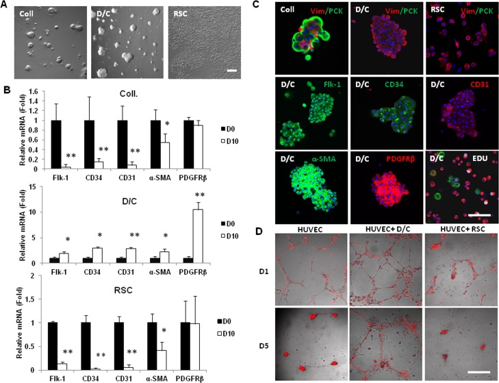
Spheres of angiogenesis progenitors in 3D Matrigel. Single cells from collagenase-isolated (Coll) clusters, D/C clusters, and limbal residual stromal cells were seeded in 3D Matrigel containing MESCM for 10 days. (A) Sphere growth was noted only from collagenase-isolated clusters (Coll) and D/C cells, but not limbal residual stromal cells. Compared to the expression level by cells immediately isolated (D0) set as 1, those of Flk-1, CD34, CD31, and α-SMA transcripts were reduced significantly in collagenase-isolated cells spheres and limbal RSC. (B) However, those of the aforementioned transcripts and PDGFRβ transcript were upregulated significantly in D/C spheres (*P < 0.05 and **P < 0.01, n = 3). (C) Collagenase-isolated cells spheres consisted of predominately pan cytokeratin (PCK)+ cells, while cells in D/C spheres and single limbal residual stromal cells were all vimentin (Vim)+. Cells in D/C spheres uniformly expressed Flk-1, CD34, CD31, α-SMA, and PDGFRβ with low EdU nuclear labeling (5%, white). (D) In 5 days co-culturing experiments on 100% Matrigel, single D10 D/C cells, but not D10 limbal residual stromal cells, stabilized the vascular network formed by human umbilical vein endothelial cells (HUVEC, prelabeled with red Q-tracker). Nuclei were counterstained by Hoechst 33342 (blue). Scale bar: 100 μm.

