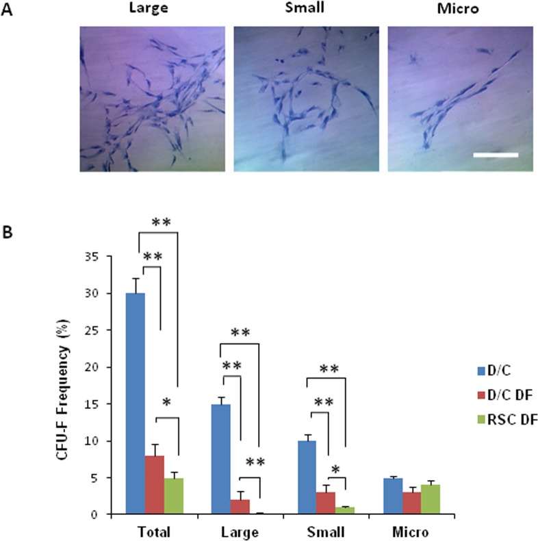
Figure 6. .
Comparison of colony-forming units-fibroblast among expanded cells. (A) After seeding at the density of 50 cells per cm2 for 12 days on plastic in DMEM with 10% FBS (DF), single cell-derived clones were stained by crystal violet. Three clones, that is large, small, and micro, were identified. (B) Colony-forming units-fibroblast (%) in D/C cells was significantly higher than those of D/C DF cells and RSC DF cells; colony-forming units-fibroblast (%) of D/C DF cells was significantly higher than that of RSC DF cells (*P < 0.05 and **P < 0.01, n = 3). Scale bar: 100 μm.

