Figure 8. .
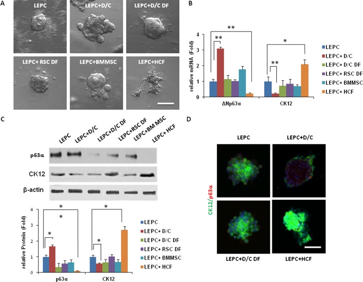
Comparison of sphere growth by reunion between limbal epithelial stem/progenitor cells and expanded cells. (A) Limbal epithelial stem/progenitor cells (LEPC) derived from dispase-isolated limbal epithelial sheets were mixed with D/C, D/C DF, and RSC DF (all at P4), as well as bone marrow-derived mesenchymal stem cells and human corneal fibroblasts to generate sphere growth on Day 10 in 3D Matrigel containing MESCM. (B) Compared to limbal epithelial stem/progenitor cells alone, expression of the ΔNp63α transcript by limbal epithelial stem/progenitor cells +D/C and limbal epithelial stem/progenitor cells + BMMSC spheres to a lesser extent was upregulated significantly, while that by limbal epithelial stem/progenitor cells + human corneal fibroblasts cells was downregulated significantly (*P < 0.05, **P < 0.01, n = 3). In contrast, expression of the cytokeratin 12 transcript was downregulated significantly in limbal epithelial stem/progenitor cells + D/C but significantly upregulated in limbal epithelial stem/progenitor cells + human corneal fibroblasts cells. The above finding of transcript expression was consistent with the protein level of p63α andcytokeratin12 based on Western blots using β-actin as a loading control (C; *P < 0.05, **P < 0.01, n = 3) and with double immunostaining between cytokeratin 12 and p63α (D).

