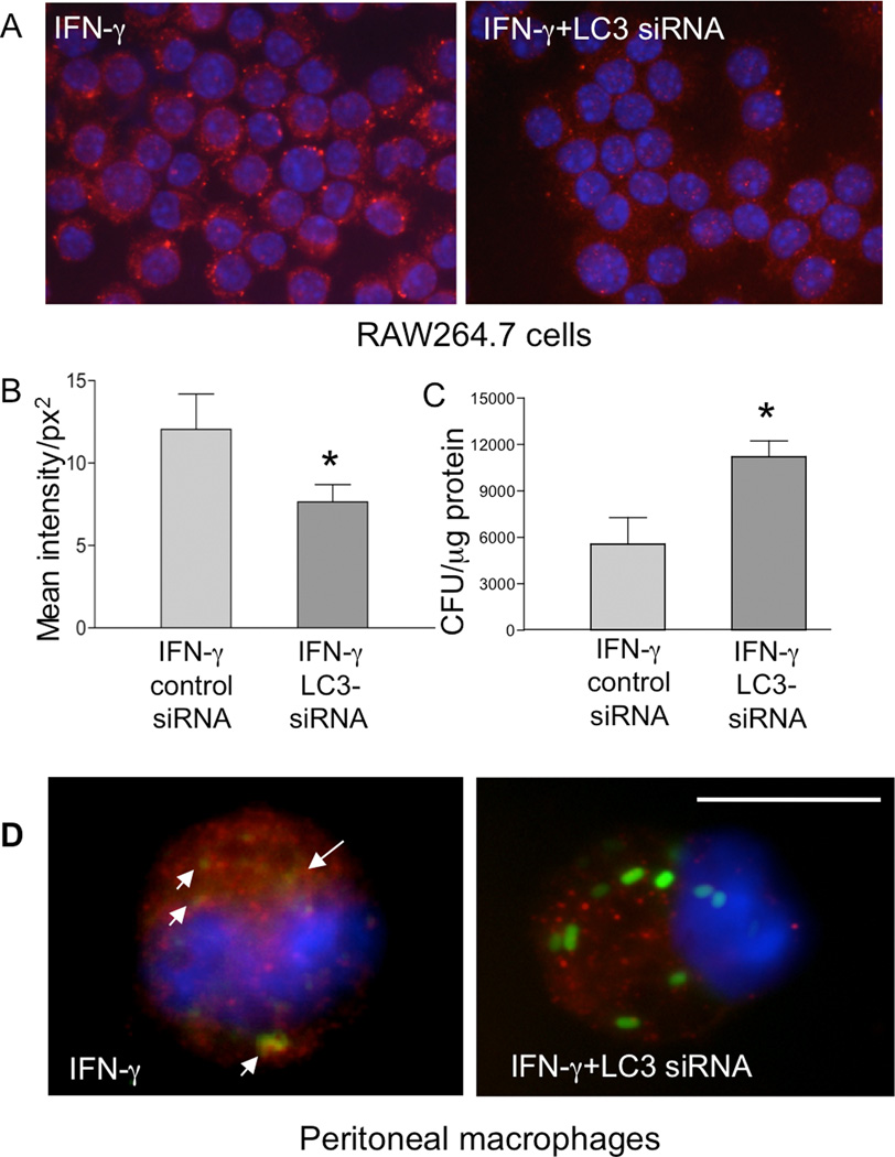
Figure 7.
siRNA-mediated knock-down of LC3 leads to impaired bacterial killing in RAW264.7 cells and peritoneal macrophages in vitro. A. RAW264.7 cells, with or without overnight IFNγ pretreatment, were transfected with or without an siRNA specific for LC3. The cells were then stained with anti-LC3 antibody as described in Figure 1. Results show that IFNγ-induced LC3 expression was effectively decreased by the siRNA (400 ×). B. Quantification of LC3 fluorescence intensity in RAW264.7 cells transfected with either a control or LC3-specific siRNA. The LC3 fluorescence intensity per cell was measured digitally using the Graph digitizing software (Nikon Imaging Software Elements). The data shown represent the mean values of fluorescence intensity obtained from 50–60 cells per slide (1 slide from each of the two experiments was examined). *p < 0.05 for a comparison of two groups. C. After knocking down LC3 expression with the siRNA, the RAW264.7 cells were used for gentamicin protection assay. The data shown represent the mean ± SD of triplicate cultures. *p < 0.05 for a comparison of two groups. D: Peritoneal macrophages were isolated from normal BALB/c mice and treated with IFNγ overnight in vitro. The cells were transfected with siRNA specific for LC3 and infected 48 h later with C. rodentium for 1 h. LC3, GFP-Citrobacter and colocalization of LC3 and bacteria were examined (1000 ×). Scale bar represents 10 µm.

