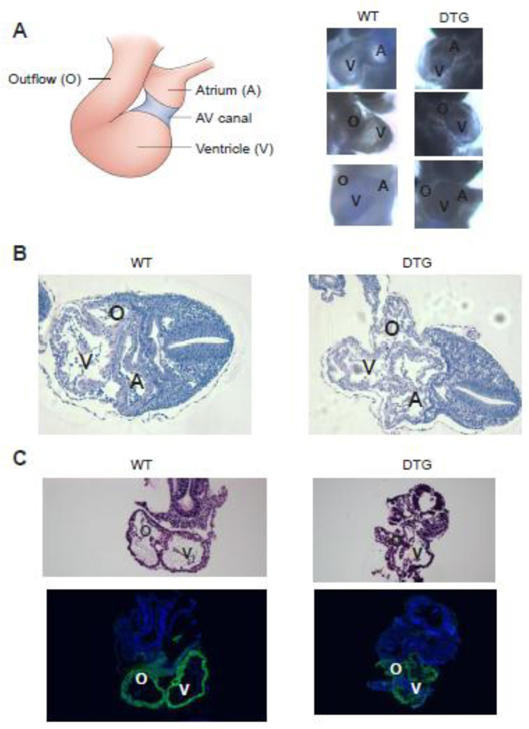
Figure 5. DTG embryos demonstrate normal looping pattern.
(A) Left: Schematic frontal view of 9.5 dpc looped heart. Right: Bright field images of 9.5 dpc WT and DTG embryos demonstrate normal looping pattern. Upper panel: left lateral view. Middle panel: right lateral view. Lower panel: frontal view. A=atrium, V=ventricle, O=outflow. (B) H&E staining of transverse sections of 9.5 dpc WT (left) and DTG embryos. Note that although looping pattern was normal, DTG embryos demonstrate collapse of all cardiac structures. A=atrium, V=ventricle, O=outflow. (C) H&E staining of sagittal sections of 9.5 dpc WT (left) and DTG. Lower panel demonstrates positive immunostaining (green) for α-smooth muscle actin (α-SMA), a marker of early cardiac mesoderm, in both WT and DTG. V=ventricle, O=outflow.

