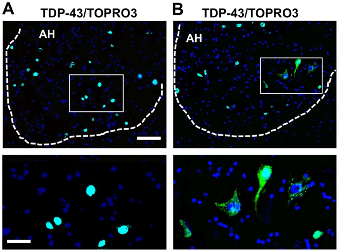
Figure 1. Abnormal localization of TDP-43 in aged mouse spinal cords.
TDP-43 (green) immunoreactivity is shown in the anterior horn (AH) cells of mouse spinal cords. (A) TDP-43 immunoreactivity (white arrows) is confined to the nuclei in the neurons, including the large motor neurons of the mouse at 6 months of age. (B) TDP-43 is absent from the nucleus and mislocalizes into the cytoplasm in the mouse at 26 months of age. Each frame area is enlarged in the inset to show the subcellular localization of TDP-43. TOPRO3 (blue) stains the nucleolus. Scale bars: 120 µm. Inset: 40 µm.

