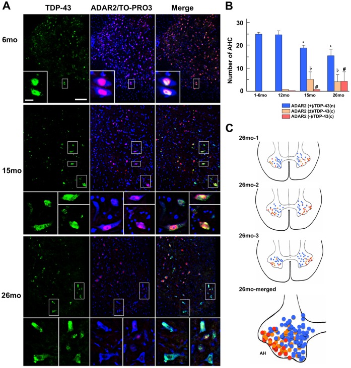
Figure 2. Motor neurons exhibiting an age-related increase of abnormal TDP-43 localization and reduced ADAR2 immunoreactivity.
(A) Representative confocal images of mouse anterior horns of the spinal cord (AH) at 6 months (6 mo), 15 months (15 mo) or 26 months (26 mo) of age. Three mice at each age were investigated. The frame area is enlarged in the inset to show TDP-43 (green) and ADAR2 (red)/TOPRO3 (blue) in the motor neurons. Both the ADAR2 and TDP-43 immunoreactivities are confined to the nuclei of all mouse motor neurons at 6 months of age. Scale bars: 100 µm. Inset: 20 µm. (B) Numbers (means ± SEMs) are shown for motor neurons with normal ADAR2- and TDP-43-immunoreactivity (blue columns), those with low ADAR2-immunoreactivity in the nucleus and mislocalization of TDP-43 (beige columns), and those that lack ADAR2-immunoreactivity in the nucleus (pink columns) in wild-type mice at different months (m) of age (1–6, 12, 15, and 26 m; n = 3 each). The number of motor neurons with normal immunoreactivity is significantly decreased (*p<0.01), whereas the number of motor neurons with abnormal ADAR2/TDP-43-immunoreactivity is significantly increased after 15 months of age compared with 12 months of age (♭ p<0.01, # p<0.01, Mann-Whitney U-test). (C) The positions of the motor neurons with abnormal immunoreactivity in the mouse AH at 26 months (n = 3). The colors (blue, beige, and pink) representing the motor neurons with different immunohistochemical patterns are the same as in Figure 2B. All of the motor neurons examined in the 3 mice at 26 months of age are plotted in the same figure as the AHs. Note that the motor neurons with abnormal immunoreactivity (beige and pink) are localized in the lateral areas of the AHs, suggesting that they are the fast fatigable motor neurons.

