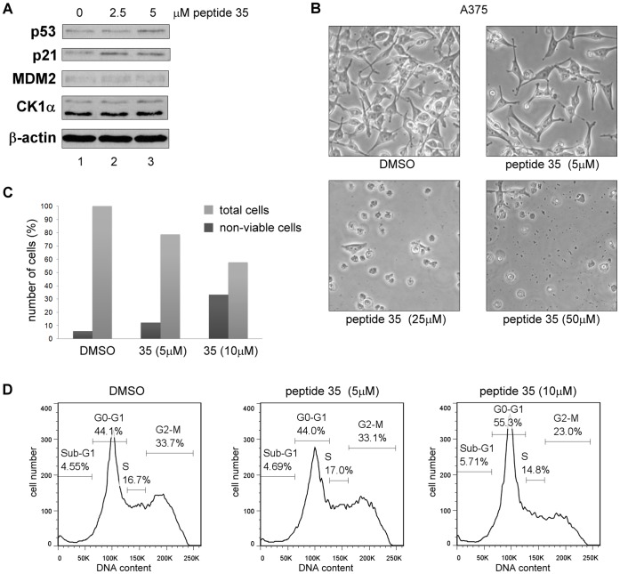
Figure 8. The CK1α peptide derived from its dominant MDM2 binding site triggers G0-G1 arrest and cell death in A375 cells.
CK1α peptide 35 was transfected into A375 cells using Nucleofectin reagent and Nucleofector device II. A DMSO control was included. (A) Protein levels were assessed 18 hours after peptide transfection by Western blotting. (B) Images of cells were captured 16 hours after transfection. (C) The number of non-viable cells was counted after treatment using Trypan Blue. Total cells are shown in percent relative to the DMSO control and percent of non-viable cells are expressed relative to each respective total cell count. (D) After treatment, the cells were harvested then fixed in ethanol followed by staining with propidium iodide. DNA content was determined by FACS and analyzed with FlowJo7 software.

