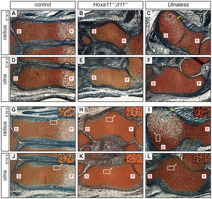
Figure 1. Lack of hypertrophic differentiation in the ulna of Hoxa11−/−;d11−/− and Ulnaless forelimbs.
Safranin-Weigert staining of E14.5 (A–F) and E16.5 (G–L) control (A, D, G, J), Hoxa11−/−;d11−/− (B, E, H, K) and Ulnaless (C, F, I, L) forelimb sections reveals disturbed chondrocyte differentiation. Hypertrophic chondrocytes are absent in ulna (E, K) and radius (B, H) of Hoxa11−/−;d11−/− mice and in the ulna (F, L) of Ulnaless mice. Only in the curve of the radius of E14.5 Ulnaless mice hypertrophic chondrocytes are detectable (C, arrow). At E16.5, mineralized matrix is produced in the radius of Ulnaless mice (I, arrow), while in the radius of Hoxa11−/−;d11−/− mice columnar chondrocytes are formed (H). These cells are not organized in columns along the longitudinal axis as in the control, but in anterior-posterior direction (G, H, see encircled columns in higher magnification of boxed regions). 160x magnification; P = proximal, D = Distal.

