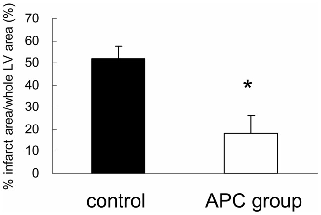
Figure 2. Assessment of myocardial infarct size after regional ischemia/reperfusion injury.
We stained slices of left ventricular (LV) tissue with hematoxylin and eosin and Sirius red 2 weeks after inducing transient ischemia. The size of the infarct area was assessed by calculating the percentage of total LV area (% infarction) using Image J. The bar graph shows that the percent infarction in the activated protein C (APC) group was significantly smaller than the controls (* p<0.05; APC versus controls).

