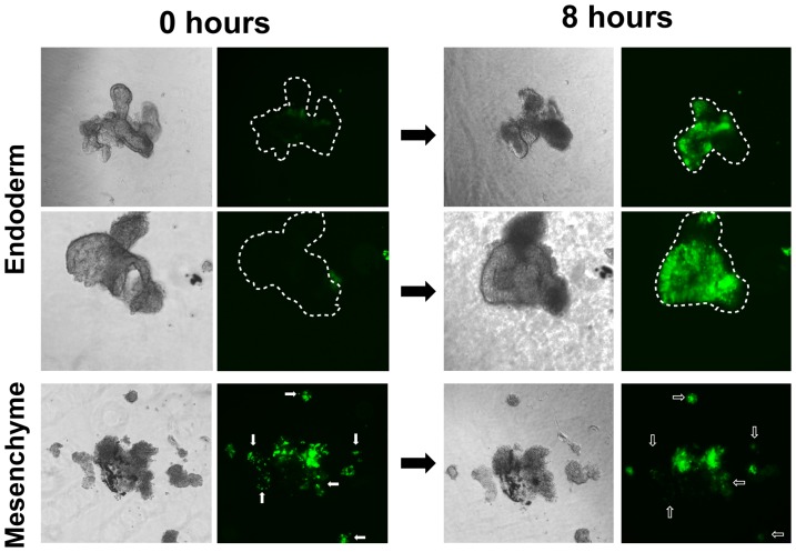
Figure 5. The BRE-eGFP reporter is activated in mesenchyme-free epithelial buds cultured in Matrigel.
Distal endodermal buds and mesenchyme were isolated by micro-dissection from E12.5 BRE-eGFP embryos and cultured for 8 hours in Matrigel. Bright field and fluorescence images from the same colonies were acquired at time 0 and 8 hours. Note the strong re-activation of the BRE-eGFP reporter in mesenchyme-free endodermal buds and the reduced (diffuse) eGFP expression in the isolated mesenchyme after 8 hours in culture. Dotted lines indicate the location of the epithelial buds in the eGFP images and small clusters of cells with reduced eGFP expression are indicated with arrows.

