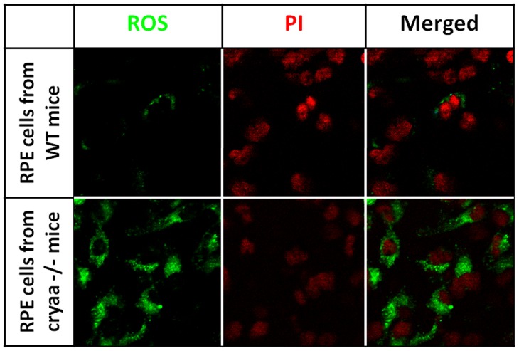
Figure 5. Increased accumulation of ROS in CRYAA −/− RPE cells.
Primary cultured RPE cells was isolated from knockout mice and treated with NaIO3. Confocal images show ROS accumulation in RPE cells. The accumulation of ROS was much stronger in RPE cells from αA-crystallin knock our mice than that from wild type mice. These results show that knock out αA-crystallin results in increased accumulation of ROS in RPE cells treated with NaIO3.

