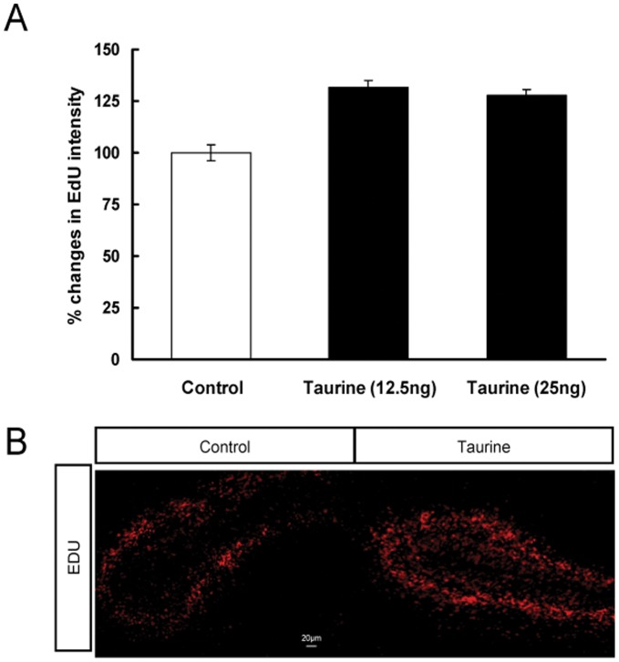
Figure 4. Effects of taurine treatment on cell proliferation in the dentate gyrus of embryonic hippocampus.
The embryonic brains were fixed at E-17. EdU intensity was measured for 6–7 identical sections per brain and at least 5–6 fetuses were used. The data of the percentage changes in EdU intensity are presented as mean ± SEM. (A). Representative images showing EdU-labeled cells (red) in the dentate gyrus of control (left panel) and taurine treated groups (right panel) (B). P>0.05, Scale bar = 20 µm.

