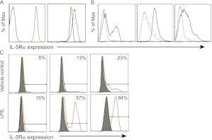
Figure 4.
Mouse neutrophils and monocytes-macrophages express the IL-5Rα in sepsis. (A) Lung and serum samples taken 18 hours after cecal ligation and puncture (CLP) were analyzed by flow cytometry for IL-5Rα, looking at receptor expression on neutrophils (Ly6 g+). The plots show IL-5Rα expression on neutrophils from a representative mouse that underwent CLP in red, and an unoperated control animal in black. Left plot represents serum neutrophils; right plot represents lung neutrophils. (B) IL-5Rα expression was examined using flow cytometry on circulating Ly6 g−CD11b+ monocytes (left panel), F4/80+ macrophages in the lung (middle panel), and F4/80+ macrophages in the spleen (right panel) 18 hours after CLP. Gray lines are unstained and isotype controls, IL-5Rα staining from a representative mouse is in black. (C) RAW264.7 mouse macrophages were stimulated with LPS, and IL-5Rα expression was examined by flow cytometry. Gray histograms are unstained controls, green is a staining control in a different channel, and red is IL-5Rα stained cells. Representative plots shown for n = 3 experiments.

