Figure 5.
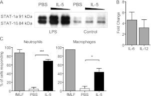
IL-5 signaling in macrophages induces activation, STAT1 phosphorylation, and cytokine production. (A and B) Primary mouse peritoneal macrophages were stimulated with LPS to induce IL-5Rα expression, followed by either 100 ng or 1 μg IL-5, or phosphate-buffered saline (PBS) control. (A) Western blot for STAT-1 performed on nuclear extracts. n = 3 experiments. (B) IL-6 or IL-12 were detected by ELISA in supernatants from IL-5–stimulated macrophages. Data are represented as fold change over vehicle-treated controls. (C) Primary mouse neutrophils or macrophages were stimulated with IL-5 and intracellular calcium changes were measured by fluorescence microscopy. fMLF is a positive control. Time-lapse images (for a total of 200 cells per experiment) were quantitated by counting the total number of cells per frame that responded to IL-5 stimulation. n = 3 experiments. Data were analyzed by Student t test; significance is defined as *P < 0.05; ***P < 0.001.

