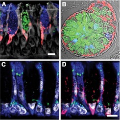
Figure 1.
AGR2 localizes to the endoplasmic reticulum (ER) of mucous cells in human airway and submucosal glands. (A) AGR2 (red) localized to cells containing MUC5AC (blue) and MUC5B (green) in airway epithelium. (B) AGR2 and mucins in submucosal glands stained as in A. (C, D) Staining with the ER marker GRP78/GRP94 (blue) and the Golgi marker giantin (green) revealed that AGR2 (red in D) is confined largely to the ER. Gray indicates autofluorescence in A, differential interference contrast in B, and nuclear (DAPI) staining in C and D. Scale bars: 5 μm.

