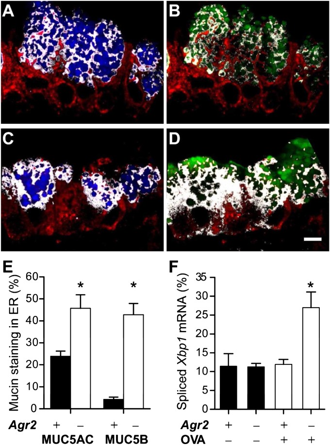
Figure 7.
Redistribution of mucins and induction of the unfolded protein response in airways of allergen-challenged AGR2-deficient mice. (A–D) ER localization of mucins in mucous cells from allergen-challenged mice. Airway epithelium from allergen-challenged wild-type control (A, B) and Agr2−/− (C, D) mice was stained for GRP78/GRP94 (A–D, red), MUC5AC (A, C, blue), and MUC5B (B, D, green). White indicates regions staining for both GRP/78/GRP94 (ER) and mucin. Scale bar: 5 μm. (E) The percentage of cellular mucin staining within the ER was determined by quantitative analysis of GRP78/GRP94 and mucin-stained images from OVA-challenged wild-type mice (closed bars, mean ± SEM for 18 total microscopic fields from six mice) and AGR2-deficient mice (open bars, 17 fields from six mice). *P < 0.05 versus wild-type mice (for the same mucin). (F) Xbp1 mRNA splicing (spliced Xbp1 mRNA amount as a percentage of total Xbp1 mRNA) was determined by PCR analysis of samples harvested by epithelial brushing from wild-type (Agr2+) and AGR2-deficient (Agr2–) mice challenged with saline (OVA–) or ovalbumin (OVA+). *P < 0.05 versus other groups.

