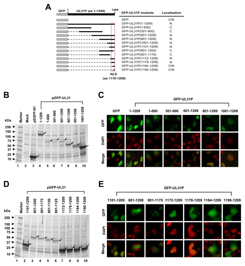
Fig. 6.
Mapping of the nuclear localization signal (NLS) of EHV-1 UL31P. Construction of GFP-UL31 fusion genes and fluorescence assays were performed as described in Materials and methods. The numbers within UL31 parentheses indicate amino acid sequences of UL31P. (A) The schematic illustrates the amino acid sequence regions of UL31P fused to the GFP and the subcellular localization of the GFP-UL31 fusion protein; N for nuclear, C for cytoplasmic, and C/N for both cytoplasmic and nuclear. (B and D) Expression of GFP-UL31 fusion proteins. The GFP fusion proteins containing truncations of UL31 ORF were detected from whole-cell lysates of transfected RK13 cells at 24 h post transfection by using an anti-rabbit GFP antibody. Arrows, bands corresponding to GFP and GFP-UL31 fusion proteins. (C and E) Localization of GFP-UL31 fusion proteins. Top, middle, and bottom panels indicate distribution of GFP-UL31 fusion protein, RK13 cell nucleus stained by DAPI, and merge of GFP and nucleus, respectively.

