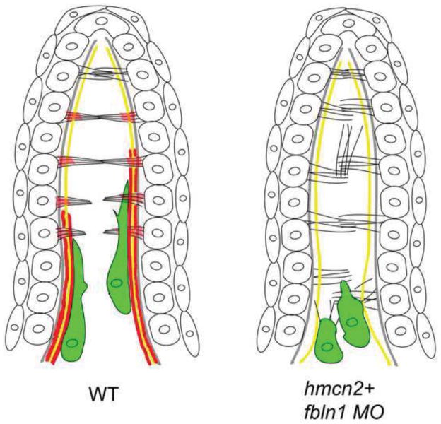
Figure 6. Simplified diagram illustrating the organization of the dorsal median fin fold in wild-type and hmcn2/fbln1 double morphant embryos at 55 hpf.
The cartoon shows the bilayered epidermal sheets, the epidermal BM in grey, actinotrichia in yellow, cross fibers in black, and fin mesenchymal cells in green. In red are the sites of Hmcn2/Fbln1 action, based on mutant analyses and anti-Hmcn2 immunohistochemistry. For simplicity, only some cross fibers are shown.
Left panel: in wild-type embryos, the cross fibers in the apical-most regions of the fin fold run in parallel and perpendicular to actinotrichia, spanning the entire interepidermal space, while in medial regions, they display a bouquet-like organization, possibly mediated by Hmcn2 located in subepidermal regions. Fin mesenchymal cells migrate on the inner surface of actinotrichia, weaving between the cross fibers. It is currently unclear whether this is directly mediated by Hmcn2 bound to actinotrichia or due to indirect effects (see Discussion).
Right panel: in the absence of Hmcn2 and Fbln1, cross fibers throughout the entire length of the fin fold remain in an organization more reminiscent of apical-most regions in wild type. The mesenchymal cells fail to migrate along the actinotrichia and remain at the base of the fin, “trapped” within cross fibers.

