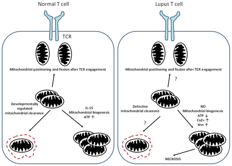
Fig. 1.
Mitochondrial homeostasis in a normal and a lupus T cells. During the formation of the IS, mitochondria are shuttled and redistributed towards the TCR signaling complex. Mitochondrial content is regulated during T cell differentiation allowing damaged mitochondria to be eliminated by autophagy. During T cell memory formation, mitochondrial biogenesis and ATP content are increased via the effect of IL-15.
However, in lupus T cell, higher mitochondrial mass and elevated potential is observed, which could be due to higher NO production in these cells. Mitochondria are larger in size and have elevated Ca2+, which alters mitochondrial movement and contributes to defective IS architecture. Removal of mitochondria could be disturbed during T cell differentiation. Contrary to the normal T cell, SLE T cells produce less ATP. The result of mitochondrial dysfunction can be necrotic cell death upon stimulation.

