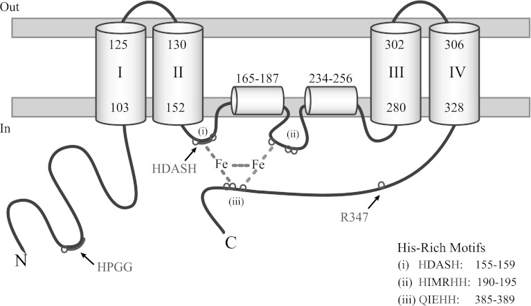
Fig. 5.
Topology model of EgΔ5D. EgΔ5D is a membrane diiron protein with three His-rich motifs. The HX(3, 4)H motif, HDASH, is located from residues 155 to 159; the HX(2, 3)HH motif, HIMRHH, located from residues 190 to 195, and the (H/Q)X(2, 3)HH motif, QIEHH, located from residues 385 to 389. EgΔ5D has four trans-membrane domains (I–IV) and two hydrophobic stretches (residues from 165 to 187, and 234 to 256). Its N-terminal cytochrome b 5 domain and C-terminal region are located in the cytoplasm

