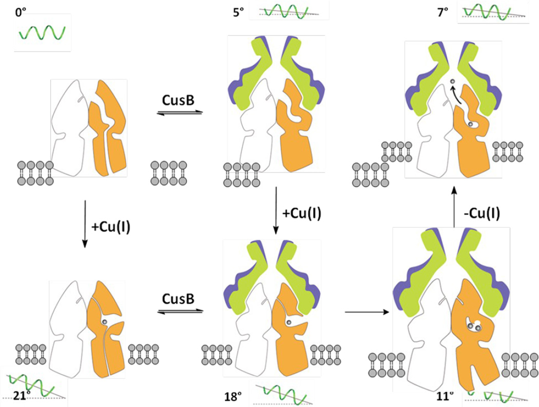
Figure 7.
Proposed model of metal ion export within the transport cycle. The model is based on different structures obtained by x-ray crystallography. Molecules 1 and 2 of CusB and a protomer of CusA are colored blue, green and orange. The angle of inclination of the horizontal helix at each state is shown in the figure. For clarity, the front protomers (one CusA and two CusB molecules) are not included.

