Fig. 1.
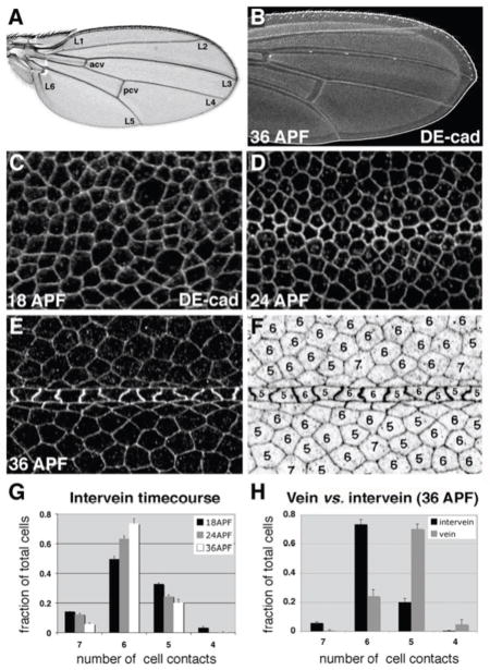
During wing epithelial cell-shape refinement, vein and intervein cells adopt different morphologies.(A) Wild-type adult wing. Longitudinal veins 1–6 (L1–L6) and two crossveins (anterior (acv) and posterior (pcv)) are labeled. (B) Pupal wing (36 h APF) labeled for DE-cadherin. (C–E) Timecourse of cell-shape refinement within the pupal wing. Wings were dissected at 18 h APF (C), 24 h APF (D), or 36 h APF (E), and labeled for DE-cad. Each image is centered on longitudinal vein L3. (F) To quantify cell shape, the number of cell-cell contacts was determined for each cell (36 h APF is shown). (G) Quantification of intervein cell shape refinement. Between 18 and 36 h APF, intervein cell shape variability decreases as hexagons become dominant (n = 4–6 images per time point, with 43–364 cells per image). (H) Quantification of vein and intervein shape differences at 36 h APF. On average, vein cells have one fewer cell-cell contact than surrounding intervein cells (n = 6 images, with 13–64 vein and 27–177 intervein cells per image). Error bars indicate SEM.

