
Fig. 8.
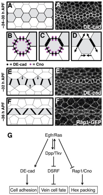
Models of adhesive changes and signaling events that determine vein–cell morphology in the wing epithelium. For schematics (A–F) shapes colored grey indicate vein-cell precursors, whereas black lines indicate DE-cad levels (thicker lines indicate higher levels of DE-cad). For representative images (A′,E′,F′), visualized proteins are indicated. (A) By ~24–30 h APF, vein precursors have narrowed to a single row of cells, which express high levels of DE-cad. (B–D) After ~24–30 h APF adhesive asymmetries arise in vein cells as Cno levels are down-regulated. (B) In hexagonal vein precursors (~24–30 h APF) there is a ~1:1 stoichiometry between DE-cad and Cno (i.e., each adherens junction is associated with a molecule of Cno). (C) Ras signaling leads to reduced levels of Cno in vein precursors, resulting in Cno-free adherens junctions that accumulate at vein-vein cell contacts. (D) Adhesive asymmetries cause vein-vein cell contacts to expand at the expense of vein-intervein regions of contact (arrows), resulting in pentagonal vein precursors (E). The row of vein precursors then straightens (arrows in E), and the 36-h APF conformation is attained (F). (G) The Egfr/Ras and Dpp/Tkv signaling pathways act in concert to specify vein-cell fate within the Drosophila wing epithelium. Egfr/Ras plays the critical role in determining the epithelial morphology of these cells, however, by regulating DE-cad and Cno in a Dpp-independent fashion.