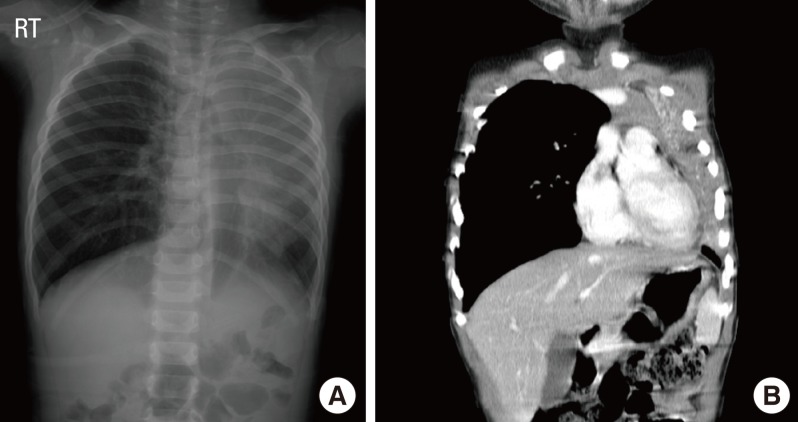
Fig. 2.
Radiographic findings of children with H1N1 pneumonia. (A) This chest X-ray shows atelectasis of the left upper lobe and consolidation in the left lower lobe. The mediastinal structure was markedly shifted toward the left side. (B) A chest CT scan demonstrates massive atelectasis of the left upper lobe, mediastinal shift to the left side, and pneumonic consolidation and pleural effusion in the left lower lobe.

