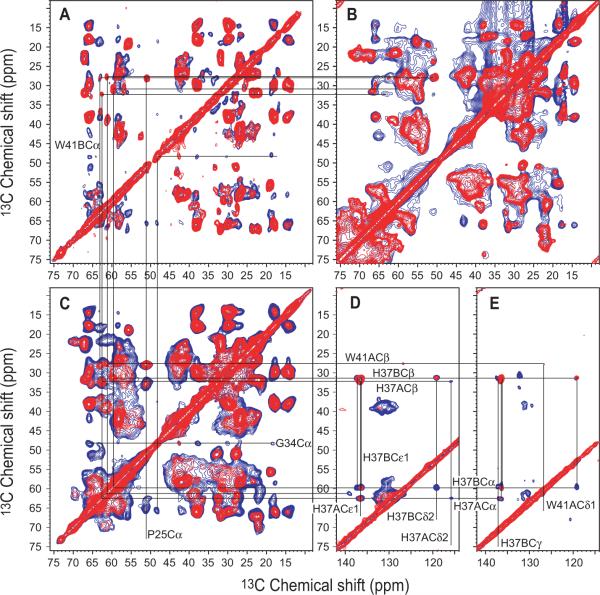
Figure 2.
13C-13C correlation spectra of uniformly [15N,13C]-labeled M2CD (A,E) and M2FL (C,D) both in reconstituted liposomes, and M2FL (B) in situ. (A,B,C) Aliphatic-aliphatic regions displaying spectra obtained with 10 (red) and 50 (blue) ms mixing times. (D,E) Aliphatic-aromatic regions displaying spectra obtained with 20 (red) and 50 (blue) ms mixing times. Lines drawn between resonances identify similar chemical shifts in the various spectra for which the sequence specific assignments have been previously achieved for M2CD. All spectra were obtained with 13C-optimized 1H/13C/15N 3.2-mm NHMFL biosolids MAS probe utilizing Low-E coil technology.[1, 2]

