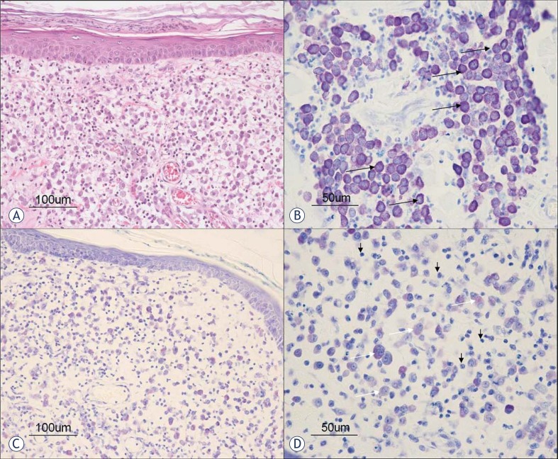
FIGURE 1.
Histological pictures of MCTs before (A and B) and after (C and D) EGT. A. Tumor mast cells are loosely arranged in the dermis without epidermal invasion (haematoxylin and eosin staining). B. Tumor mast cells (arrow) have a well granulated metachromatic cytoplasm (toluidine blue staining). C. Decreased number of mast cells in the dermis after the treatement (toluidine blue staining). D. Note the degranulated tumor mast cells with metachromatically weakly stained cytoplasm (white arrows) intermingled with numerous inflammatory cells (black arrows) (toluidine blue staining).

