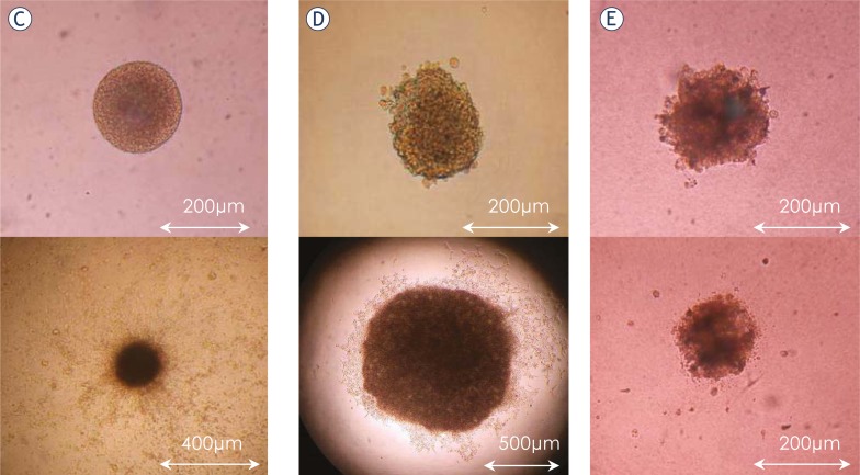
FIGURE 4.
3D spheroid invasion assay. Spheroids were imbedded into collagen I and the invasion distance (panel A) and diameter (panel B) measured under the light microscope for up to 21 days. The average invasion distance was significantly higher (p<0.05) for spheres of NNSC than for spheres of NCH644. The average spheroid size did not change significantly in NNSC, but increased (p=0.009) in spheres of GBM stem cells, NCH644. GBM biopsy spheroids did not change in size and very few cells invaded the surrounding collagen. Panels C, D and E show the spheroids of NNCS, NCH644 cells and GBM spheroid, respectively at the start (upper panel) and after 21 days (lower panel) of the experiment.


