Fig. 6.

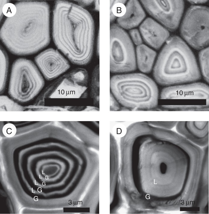
(A, B) Ultraviolet micrographs of transverse sections of phloem fibres in Mallotus japonicus collected in December, taken at 280 nm. (C, D) Confocal images of phloem fibres stained with acriflavine. (A, C) Tension side; (B, D) opposite side.

(A, B) Ultraviolet micrographs of transverse sections of phloem fibres in Mallotus japonicus collected in December, taken at 280 nm. (C, D) Confocal images of phloem fibres stained with acriflavine. (A, C) Tension side; (B, D) opposite side.