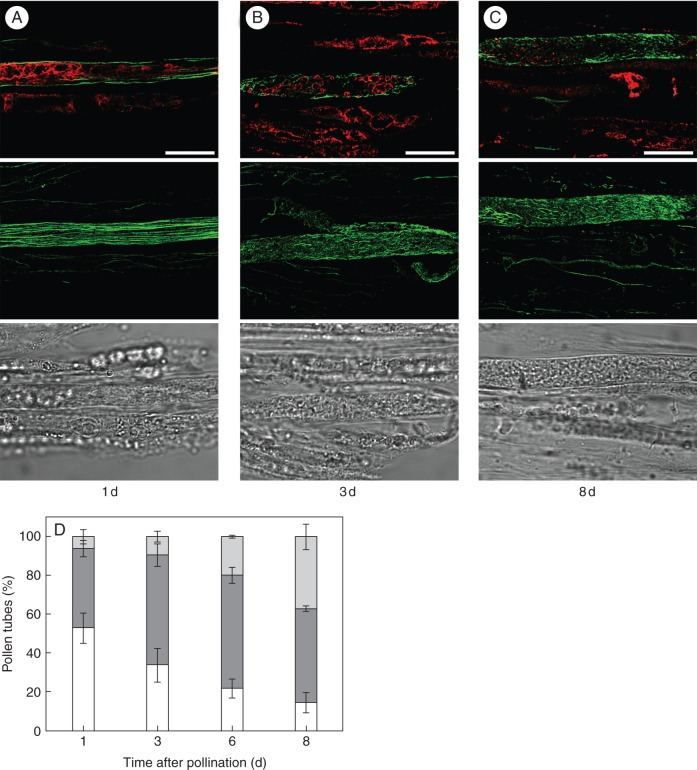
Fig. 4.
Disorganization of F-actin cytoskeleton precedes vacuolar breakdown in incompatible pollen tubes during the in vivo SI response. Colocalization of F-actin (green) and vPPase (red) in incompatibly pollinated pistils. The images are representative of the three patterns observed: (A) organized F-actin and vacuolar endomembrane system; (B) disorganized F-actin and organized vacuolar endomembrane system; (C) disorganized F-actin and vacuolar endomembrane system. Merged optical sections (top panels), full projections of phalloidin-stained pollen tubes (central panels) and the corresponding bright fields (bottom panels) are shown. The days after pollination are indicated below the images. Scale bar = 10 µm. (D) Quantification of morphology of F-actin and vacuolar endomembrane system. White, organized F-actin and vacuolar system; dark grey, disorganized F-actin and organized vacuolar system; light grey, disorganized F-actin and vacuolar system. Seventy-five pollen tubes were examined in each experiment. The values are the average ± s.d. for two or more independent experiments.

