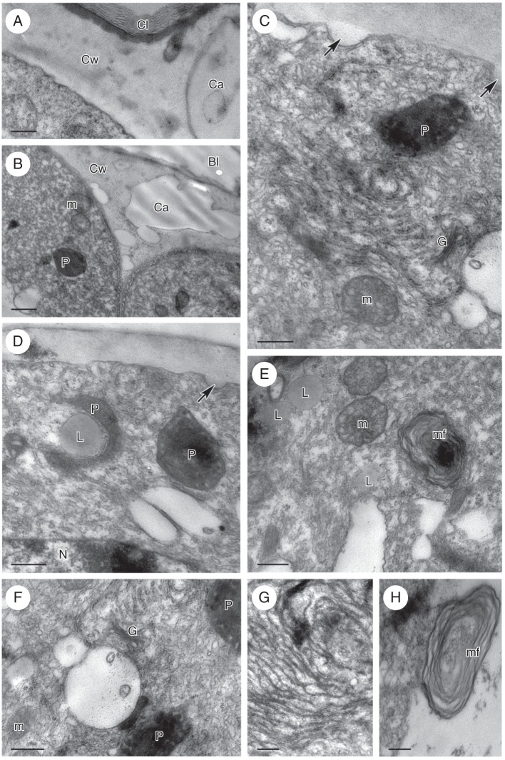
Fig. 5.
Zygostates grandiflora, TEM. (A) Cell wall with lamellate cuticle, beneath which occur an osmiophilic layer and a large wall cavity. (B) Cell wall cavities and blistered cuticle. The parietal cytoplasm contains abundant ER profiles, plastids and mitochondria. (C) Small protuberances or ingrowths of cell wall (arrows). The cytoplasm contains plastids, dictyosomes (Golgi apparatus), mitochondria and ER that is in close contact with the plasmalemma. (D) Lipid droplet associated with plastid, whereas the ER is continuous with the plasmalemma. Arrow indicates protuberance or ingrowth of cell wall. (E) Profiles of ER, mitochondria, lipid bodies and cytoplasmic, myelin-like figure. (F) Plastids, dictyosomes (Golgi apparatus), small vacuoles and predominantly smooth ER with dilated cisternae. (G) Detail of myelin-like figure formed in the cytoplasm from ER membranes. (H) Myelin-like figure extruding into vacuole. Scale bars: (A, C, E–H) = 0·5 µm; (B, D) = 1 µm. See Fig. 1 for abbreviations.

